
The Liver–Skin Connection in Dogs
- Fruzsina Moricz

- Apr 3
- 11 min read
About 70% of a dog’s body protein is constantly being remodeled in the liver and skin. When the liver can’t keep up with that work, one of the first places it quietly shows is not in the stool, or the eyes, but on the surface: the coat, the pads, the ears, the muzzle.
That’s the core of the liver–skin connection in dogs: a metabolic organ deep in the abdomen creating very visible, very tangible problems on the outside.
If you’ve watched your dog’s skin crack and crust, or their once-glossy coat turn dull and sparse while blood tests talk about “liver enzymes,” it can feel like two separate problems. They aren’t. They’re often the same story, told in different tissues.

This article walks through that story—what we know, what we don’t, and how to think about chronic skin issues when the liver might be involved.
Why a sick liver can show up as bad skin
The liver is not just a “filter.” It is a manufacturing plant. For skin and coat health, it is responsible for:
Making many of the proteins that hold the skin together
Processing and distributing fatty acids that keep the skin barrier flexible and the coat shiny
Storing and activating vitamins and trace minerals (like zinc) used in skin repair
Managing glucose and other fuels that skin cells need to grow and renew
When the liver is damaged—by chronic disease, endocrine problems, toxins, infection, or tumors—its ability to do all of this can drop. The result is a kind of systemic malnutrition, even if the dog is eating a good diet.
The skin, being a fast‑turnover, metabolically hungry organ, is one of the first places that “shortage” shows up.
In mild or early disease, that might look like:
Dull, dry coat
Slow hair regrowth after clipping
Flaky skin (dandruff)
Recurrent skin infections
In more advanced or specific liver disorders, the skin can develop very characteristic lesions that are painful and hard to ignore. This is where hepatocutaneous syndrome comes in.
Hepatocutaneous syndrome: when liver disease writes on the skin
You may hear several different names for essentially the same clinical picture:
Hepatocutaneous syndrome (HCS) – emphasizes the liver–skin link
Superficial necrolytic dermatitis (SND) – what the skin biopsy looks like
Necrolytic migratory erythema (NME) – a term borrowed from human medicine
They all describe a pattern where chronic liver dysfunction (or sometimes severe metabolic disease like uncontrolled diabetes) leads to striking, painful skin lesions.
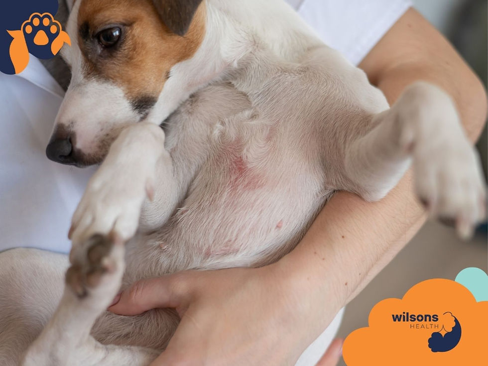
What it looks like on the dog
Hepatocutaneous syndrome tends to appear in older dogs and has some favorite locations:
Footpads – thickened, cracked, crusted pads; can split and bleed
Muzzle and lips – red, crusty, sometimes ulcerated
Around the eyes – scaling, redness, crusting
Pressure points and genital area – erosions, ulcers, crusts
Owners often describe:
Reluctance to walk or jump because the pads are so sore
A foul smell from infected, oozing skin
A dog that seems “miserable in their own body”
Under the microscope, the skin shows epidermal necrosis (superficial layers of skin cells dying), separation, and thick crusts—hence “superficial necrolytic dermatitis.”
Under the ultrasound probe or biopsy needle, the liver often shows:
Nodular changes
Fibrosis (scar tissue)
Hepatocellular necrosis (liver cells dying)
Sometimes masses or evidence of metabolic liver disease
These are not cosmetic issues. They are the skin’s response to a body-wide metabolic imbalance.
The metabolic triangle: liver, hormones, and skin
Hepatocutaneous syndrome almost never exists in isolation. It often sits in the middle of a triangle:
Liver disease (fibrosis, nodular hyperplasia, tumors, chronic hepatitis)
Endocrine disease (especially diabetes mellitus, sometimes Cushing’s disease)
Severe protein and amino acid deficiency at the tissue level
Even when blood protein levels look “normal,” the skin may not be getting the building blocks it needs. Research and case series suggest that:
The syndrome is linked to metabolic derangements and protein deficiencies rather than a single toxin or pathogen [2][5].
Managing underlying endocrine conditions (like diabetes or Cushing’s) can sometimes improve the skin lesions [2][5].
Treatment protocols that include intravenous amino acid and lipid infusions can lead to partial or complete remission of the skin lesions, even if the liver does not fully recover [5].
One study reported that some dogs needed only 1–3 IV infusions to see major skin improvement, while another needed 13 separate infusions to reach remission [5]. That variability is part of what makes this condition emotionally and practically challenging.
When infection links liver and skin: the leishmaniasis example
The liver–skin connection isn’t limited to metabolic and endocrine disease. Infectious diseases can also tie them together.
In canine visceral leishmaniasis, a parasitic disease prevalent in some regions, researchers have found:
Dogs often show chronic dry desquamation (scaling) especially on the ears and limbs [1].
There is a strong positive correlation between parasite load and hepatic collagen deposition (a measure of liver fibrosis):
r = 0.7124, P < 0.0001 [1].
The more fibrosis in the liver, the more likely and more severe the skin lesions.
Put simply: as the parasite damages the liver, the liver responds with fibrosis, and that internal scarring shows up externally as skin disease.
This reinforces a key idea:Skin problems can be a visible marker of invisible liver damage.
Breeds, faces, and hidden liver strain
You might read that hepatocutaneous syndrome is more common in certain breeds:
Miniature Poodles
Lhasa Apsos
Cocker Spaniels
Current thinking is cautious: this could reflect a true genetic predisposition or simply referral bias—these breeds being overrepresented in specialty clinics [2]. Large, robust dogs can and do develop the same syndrome.
Another emerging piece comes from liver elastography—a way to measure how stiff the liver is, similar to checking how firm a fruit feels.
Studies in brachycephalic dogs (short-nosed breeds like Bulldogs and Pugs) with brachycephalic obstructive airway syndrome (BOAS) found:
These dogs have significantly higher liver stiffness on elastography compared to non‑brachycephalic dogs [3].
That suggests a chronic, inflammatory, or congestive environment in the liver, even before classic “liver disease” is diagnosed.
We don’t yet have direct proof that this increased liver stiffness translates into specific skin problems. But it suggests that some dogs may carry silent liver strain for years, and their skin and coat may be among the first things to show that something is off.
How veterinarians connect the dots: from coat to liver
When a dog presents with chronic, unusual, or painful skin problems, especially in older age, a thoughtful vet will often step back and ask:
“Is this just a skin problem, or is the skin reporting on something systemic?”
The work‑up may include several layers.
1. History and physical exam
Clues that push the vet toward liver involvement:
Age (hepatocutaneous syndrome is rare but mostly seen in older dogs [2][5])
Distribution of lesions: pads, muzzle, around the eyes, genital area, pressure points
Signs of systemic disease:
Drinking and urinating more
Weight loss or muscle wasting
Lethargy, reduced stamina
Neurologic changes (in severe liver disease)
Breed (e.g., Miniature Poodle, Lhasa Apso, Cocker Spaniel) – not diagnostic, but raises suspicion [2]
2. Bloodwork and liver enzymes
Common tests include:
ALT (alanine aminotransferase)
AST (aspartate aminotransferase)
ALP (alkaline phosphatase)
GGT (gamma‑glutamyl transferase)
These are biomarkers of liver cell damage or bile flow issues [4]. In many dogs with liver‑related skin disease, they are elevated.
But there’s an important nuance:There is significant overlap in enzyme levels between healthy and diseased dogs. High enzymes mean “something is happening,” not “we know exactly what and how bad.”
Your vet may also check:
Bile acids – how well the liver processes and recycles bile
Glucose – screening for diabetes
Cholesterol and triglycerides
Albumin and total protein
Electrolytes and other organ markers
3. Imaging: looking at the liver itself
Because enzymes can only say so much, imaging is often the next step:
Ultrasound – to look for:
Nodules, masses, or generalized changes in liver texture
Fibrosis patterns
Abnormal blood vessels
Radiographs (X‑rays) – to see liver size and shape, and look for masses
CT scans – in complex cases, to better define masses or plan surgery [4]
In some reported cases, pedunculated liver masses (tumors hanging from the liver on a stalk) have been associated with skin manifestations [4]. Without imaging, those would be completely invisible.
4. Biopsy: the ethical crossroads
A liver biopsy—either via ultrasound guidance or surgery—can give the most definitive information:
Type of liver disease (inflammatory, fibrotic, neoplastic, metabolic)
Severity and pattern of fibrosis
Prognostic clues
But biopsies are invasive and carry risks, especially in dogs who may have:
Clotting abnormalities
Poor general condition
Advanced age
This creates a genuine ethical tension:
Diagnostic clarity vs. procedural risk
More information vs. the stress and cost of getting it
Different vets and owners make different choices here, and there isn’t always a single “right” answer.
Treatment: what can actually help?
There is no one‑size‑fits‑all protocol for liver‑related skin disease. Management usually has several strands woven together.
1. Addressing the underlying disease
Depending on the diagnosis, this may include:
Endocrine management
Insulin therapy for diabetes mellitus
Treatment for Cushing’s disease
Improvement in metabolic balance can translate into better skin [2][5].
Treatment for infectious disease
For conditions like leishmaniasis, antiparasitic therapy aims to reduce parasite load, which in turn can reduce liver fibrosis and skin signs [1].
Surgery or oncology care
For liver masses or tumors, if appropriate and feasible [4].
These decisions are individualized and should be made in close consultation with your vet or an internal medicine specialist.
2. Nutritional and metabolic support
Because protein and amino acid deficiency at the tissue level is central in hepatocutaneous syndrome, many treatment plans focus on:
High‑quality, highly digestible protein sources (within what the liver can tolerate)
Supplemental amino acids, often via:
Intravenous amino acid infusions
Sometimes combined with lipid (fat) infusions [5]
Essential fatty acids for skin barrier support
Micronutrients such as zinc and vitamins, where indicated
In published cases:
Some dogs showed dramatic improvement in skin lesions after only a few amino acid/lipid infusions.
Others required many weeks and repeated treatments—one dog needed 13 infusions before remission [5].
Importantly, skin improvement did not always mean the liver was “fixed.” The underlying liver disease often remained chronic or progressive.
This is where expectation‑setting becomes crucial:The goal is often comfort and quality of life, not a cure.
3. Local skin care and pain management
Even as you and your vet work on the liver and metabolism, the skin itself needs care:
Gentle cleansing of crusted, ulcerated areas
Managing secondary infections
Protecting cracked pads
Pain control so the dog can walk, sleep, and interact more normally
This can feel like “just symptomatic treatment,” but for the dog, symptom relief is the difference between suffering and tolerable daily life.
The emotional reality: living with a dog whose skin shows their liver disease
Chronic liver‑related skin disease is not just medically complex; it’s emotionally heavy.
Owners often describe:
Guilt – for not noticing earlier, or for struggling to afford intensive treatments
Helplessness – when treatments give inconsistent or temporary results
Exhaustion – from frequent vet visits, complex medication schedules, and home care
Anxiety about prognosis – not knowing if you’re buying months, a year, or more
Veterinarians, on the other side of the exam table, feel their own tensions:
Wanting to be honest about prognostic uncertainty without taking away hope
Balancing invasive diagnostics against the dog’s comfort and the owner’s resources
Feeling frustration when advanced treatments yield only partial or short‑lived results
Navigating euthanasia timing when the dog’s skin is clearly suffering but other systems seem “okay”
Acknowledging this emotional landscape doesn’t solve it, but it can make it less lonely. You are not the only person to feel overwhelmed by a dog’s skin and liver disease.
How to think about “next steps” if you suspect a liver–skin link
You don’t need to arrive at your vet’s office with a self‑diagnosis. But having a mental map can make the conversation more productive and less frightening.
Questions you might bring to your veterinarian
“Could these skin changes be related to her liver or another internal disease?”
“Would liver enzymes or bile acids help us here?”
“If her liver enzymes are high, what are the likely causes in a dog like her?”
“What might ultrasound or other imaging add to the picture?”
“If we consider a liver biopsy, what are the risks and what would we gain in terms of treatment decisions?”
“If this is hepatocutaneous syndrome, what are the realistic goals—comfort, time, remission?”
“What signs would tell us that treatment is helping? What signs would tell us the disease is progressing?”
Ways to frame decisions for yourself
Instead of “Should we do everything?” it can help to ask:
What burden does each test or treatment place on my dog (pain, fear, travel)?
What burden does it place on my household (time, finances, emotional energy)?
What benefit—in comfort, function, or clarity—am I hoping for from each step?
What does quality of life look like for my particular dog?
Sometimes, a dog and family are well‑suited to frequent hospital visits and aggressive metabolic support. Sometimes, the kinder path is a simpler, palliative plan focused on pain relief, gentle skin care, and shared time.
Both can be deeply loving choices.
What we know solidly vs. what we’re still figuring out
It may help to see the science in two columns:
Well‑established | Emerging / Uncertain |
Liver dysfunction can cause distinctive skin lesions in dogs, especially hepatocutaneous syndrome / superficial necrolytic dermatitis [2][5]. | The exact biochemical pathways that lead from liver damage to these specific skin lesions in dogs are still being mapped; much is extrapolated from human medicine [2]. |
In diseases like leishmaniasis, there is a positive correlation between liver fibrosis and skin manifestations [1]. | Whether we can use elastography (liver stiffness measurements) as a routine screening tool for liver involvement in systemic diseases like BOAS is not yet clear [3]. |
Amino acid and lipid infusions, along with supplementation, can improve or temporarily resolve skin lesions in many dogs with hepatocutaneous syndrome, though responses vary widely [5]. | The long‑term outcomes of combined therapies for hepatocutaneous syndrome—who does best, for how long, and why—are still being clarified [5]. |
Diagnosing liver masses or diffuse liver disease can be challenging but is improved by ultrasound, CT, and sometimes biopsy [4]. | The role of genetics and breed predisposition in hepatocutaneous syndrome needs stronger epidemiological data [2]. |
Uncertainty is not failure; it’s the honest state of a field that is still growing. Knowing where the edges are can make it easier to tolerate the parts of your dog’s care that feel like “trial and adjustment” rather than precision.
A brief glossary for your notes
You may hear these terms; it can help to have them in one place.
Hepatocutaneous Syndrome (HCS): A condition where chronic liver dysfunction leads to characteristic, often painful skin lesions, usually in older dogs. Frequently associated with metabolic derangements and tissue‑level protein deficiencies [2][5].
Superficial Necrolytic Dermatitis (SND): The skin pathology seen in HCS: superficial layers of the skin die and separate, leading to erosions, ulcerations, and thick crusts [2][5].
Necrolytic Migratory Erythema (NME): A descriptive term for similar skin lesions in humans; sometimes applied to the canine condition.
Liver Fibrosis / Collagen Deposition: Build‑up of scar‑like tissue in the liver’s extracellular matrix. In diseases like leishmaniasis, more fibrosis is associated with more severe skin lesions [1].
Liver Enzymes (ALT, AST, ALP, GGT): Blood markers that often increase when liver cells are damaged or bile flow is impaired. Helpful but not definitive; they must be interpreted in context [4].
Elastography: An imaging technique that measures liver stiffness. Brachycephalic dogs with BOAS have been shown to have stiffer livers on elastography, suggesting systemic effects of their airway disease [3].
Living with the connection
The liver–skin connection can feel unfair. Your dog’s skin becomes the billboard for an internal problem that is harder to see and harder to fix.
Yet there is also a strange kind of gift in it:Because the skin is visible, it can act as an early‑warning system and a real‑time gauge of how well the body is coping.
When the pads soften, the crusts thin, the coat regains a bit of shine after treatment—that is not just “cosmetic improvement.” It is your dog’s metabolism responding, however partially, to the support you and your veterinary team are giving.
And when improvement doesn’t come, or doesn’t last, that too is information: about limits, about progression, about when comfort might matter more than control.
Understanding the biology doesn’t make the decisions easy. But it can make them clearer—and clarity, in long‑term caregiving, is often the kindest thing we can offer ourselves.
References
Silva FL, et al. “Liver extracellular matrix remodeling in canine visceral leishmaniasis and its relationship with skin lesions.” PLoS One. 2009;4(9):e7465. (PMC2768152).
Outerbridge CA. “Hepatocutaneous syndrome: When liver dysfunction affects skin.” dvm360. Available at: https://www.dvm360.com/.
Sura PA, et al. “Liver and spleen elastography in brachycephalic dogs with obstructive airway syndrome.” Scientific Reports. Nature Publishing Group. Available at: https://www.nature.com/.
Sato T, et al. “Pedunculated liver masses causing clinical signs in dogs: a case report.” Frontiers in Veterinary Science. Available at: https://www.frontiersin.org/.
Marchetti V, et al. “Treatment and outcomes of dogs with hepatocutaneous syndrome and superficial necrolytic dermatitis.” BMC Veterinary Research. 2022;18:41. (PMC8783367).
Hobi S, et al. “Diagnosis and treatment of hepatocutaneous syndrome in a Maltese dog.” Journal of Small Animal Practice. British Veterinary Association Journals (bvajournals).


Comments