
The Four Phases of Wound Healing in Dogs
- Fruzsina Moricz

- 1 day ago
- 10 min read
A dog’s surgical incision can reach about 80% of its original strength—yet it may take months or even years to get there.[9][11] In the first week it can look pink and bumpy, then suddenly angry and swollen, then oddly shiny as new skin creeps across. To human eyes, that can feel like “better, then worse, then maybe infected?”
Biologically, though, this is often exactly what normal wound healing looks like.
Understanding the four phases of wound healing in dogs—what’s supposed to happen and when—can turn a very stressful, guilt‑tinged experience into something more predictable and less frightening. You’re not guessing; you’re watching a process unfold.
The four phases at a glance
Veterinary medicine describes four classic, overlapping phases of wound healing in dogs:
Inflammation (roughly Day 0–3)
Debridement (overlaps with inflammation)
Repair / Proliferation (about Day 3–14)
Maturation / Remodeling (weeks to months, sometimes longer)
They don’t switch on and off like light switches. They blend into each other, and the exact timing depends on:
The type of wound (surgical incision vs. bite wound vs. chronic sore)
Location (high‑motion areas like joints tend to be slower)
Your dog’s age, general health, and underlying diseases
Infection, trauma, or repeated licking/chewing
Think of these phases less as four neat boxes and more as four “jobs” the body must complete: stop the bleeding, clean up, rebuild, then reinforce.
Phase 1: Inflammation – the controlled emergency (Day 0–3)
What’s happening biologically
Right after injury or surgery, your dog’s body goes into controlled crisis mode:
Hemostasis (stopping bleeding):
Blood vessels briefly tighten (vasoconstriction).
Platelets rush in and form a clot, creating a temporary plug.
Chemical alarm:
Damaged cells release signaling molecules that call in immune cells.
Immune cell arrival:
Neutrophils are the first responders, targeting bacteria and debris.
Macrophages arrive a bit later, continuing cleanup and releasing growth factors that tell the body, “Start building.”
What you might see
Redness around the wound
Warmth to the touch
Swelling
Some pain or sensitivity
Clear to slightly blood‑tinged fluid on dressings
These are the classic signs of inflammation—and in the first couple of days, they are not automatically signs of “something’s wrong.” They’re signs the body has noticed the problem and is mobilizing.
Where debridement fits in
Debridement—removal of dead or contaminated tissue—starts here and overlaps with the inflammatory phase.
White blood cells begin autolytic debridement: they enzymatically break down dead tissue and bacteria while mostly sparing healthy tissue.[1][7][12]
In some wounds, your vet may assist with surgical or mechanical debridement to give the body a cleaner starting point.
Owner experience in this phase
This is often the “panic phase” for humans:
The wound looks dramatic.
Your dog may guard the area or seem uncomfortable.
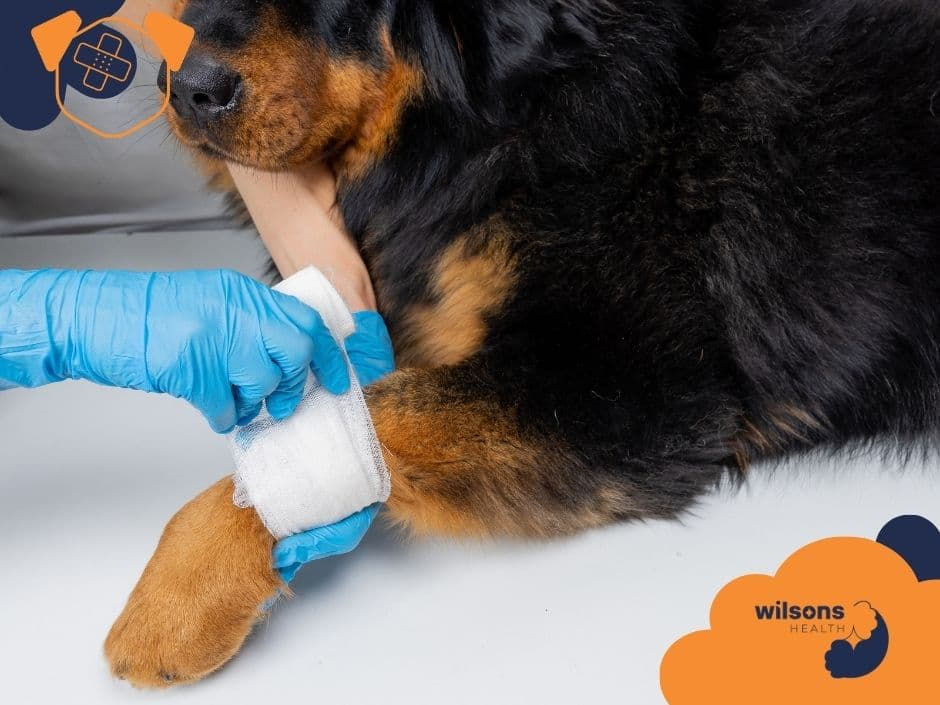
Bandages get stained, and it’s hard to tell what’s “normal” fluid vs. infection.
What helps:
Knowing that some redness, warmth, and swelling in the first 48–72 hours is expected.
Having clear instructions from your vet on what is not okay (e.g., foul odor, thick green/yellow discharge, severe pain, or sudden worsening after initial improvement).
Taking a photo once a day to track changes rather than relying on memory.
Phase 2: Debridement – the cleanup crew (overlaps Days 1–5+)
Debridement doesn’t get its own dramatic “look” the way inflammation does, but it’s crucial, especially in messy or chronic wounds.
What’s happening biologically
Neutrophils and macrophages continue to digest and remove:
Necrotic (dead) tissue
Bacteria
Foreign materials (dirt, hair, etc.)
This is highly selective when it’s autolytic: the body aims to clear only what’s damaged and keep what can still function.[1][7][12]
In chronic or heavily contaminated wounds, this phase can be prolonged and may require repeated veterinary debridement. Until the wound bed is reasonably clean, the body is reluctant to move fully into rebuilding mode.
What you might see
The wound may still look “messy,” with slough (soft, yellowish tissue) or patches of dead tissue early on.
Over time, you may notice:
Less obviously dead tissue
A cleaner, more uniformly moist surface
Less foul odor (if present initially)
Why vets care so much about this phase
A well‑debrided wound:
Is less likely to become chronically infected
Responds better to dressings and advanced therapies
Transitions more smoothly into the repair phase
This is why you’ll hear so much about wound cleaning, appropriate moisture, and infection control in the early days.[11][12][13] These aren’t cosmetic steps; they’re how we give the body a fair chance to move on to rebuilding.
Phase 3: Repair / Proliferation – the rebuilding phase (Days 3–14)
Once bleeding is controlled and the worst of the debris is cleared, the body starts building new tissue. This is where things often look “better, then worse, then better again” to owners.
What’s happening biologically
This phase has three big jobs:
Granulation tissue formation
Fibroblasts move into the wound and begin laying down collagen, the protein scaffolding of new tissue.
New blood vessels grow in (angiogenesis), giving the wound a pink, moist, bumpy appearance.
This pink tissue is called granulation tissue—it’s a very good sign.[1][3][5][7][9][11][12]
Contraction
The wound edges gradually pull toward each other as specialized cells contract, like tiny internal drawstrings.
Epithelialization
Epithelial cells (skin surface cells) migrate from the edges and sometimes hair follicles to cover the wound with new skin.
Initially, this new skin may look thin, shiny, or slightly different in color.
What you might see
A moist, pink to red, slightly bumpy surface filling in an open wound
Edges that look less sharp and more “rounded in” as they contract
Gradual reduction in wound size
Less fluid over time, often changing from bloody to clearer
Owners sometimes worry that granulation tissue looks “too red” or “angry.” In many cases, this bright, healthy, moist tissue is exactly what we want. Your vet will distinguish between healthy granulation and problematic overgrowth or infection.
Advanced therapies often target this phase
Research in dogs has focused heavily on helping this phase go faster or more completely, especially in chronic or large wounds:
Stem cell therapy (allogeneic adipose‑derived mesenchymal stem cells, ASCs):
In a controlled study of 24 dogs, ASC‑treated wounds showed significantly improved contraction and re‑epithelialization compared to conventional treatment.
At 90 days, ASC‑treated wounds had over 97% re‑epithelialization, outperforming standard care.[2]
Amniotic membrane (AM) dressings:
Both fresh and lyophilized (freeze‑dried) bovine AM accelerated healing of full‑thickness skin wounds.
Wounds treated with AM had closure rates of 88.6% at week 3 and 100% at week 5, compared to 55.7% and 84% in controls.[4]
Lyophilized AM in particular led to faster closure and narrower wound edges at 5 weeks.
Platelet‑rich fibrin (PRF):
In a case series of 8 dogs with naturally occurring wounds, autologous PRF (made from the dog’s own blood) enhanced granulation and sped wound closure.[6]
Laser therapy (photobiomodulation):
Studies suggest certain laser protocols can stimulate cellular activity and accelerate incision healing in dogs.[8]
These therapies don’t replace the fundamental phases of healing—they attempt to support or accelerate them, especially in wounds that are stuck or slow.
What’s still uncertain
The best protocols (dose, frequency, type of stem cells or PRF)
How the long‑term tissue quality compares to standard healing[2][4][6][8]
Which dogs benefit most and when the cost is justified
If your vet mentions these options, it’s reasonable to ask:
“Which phase of healing are we trying to help?”
“What’s the evidence in dogs like mine?”
“What are the realistic goals—faster closure, better tissue quality, less scarring?”
Phase 4: Maturation / Remodeling – the long, quiet phase (weeks to months+)
By the time most owners feel emotionally “done” with a wound, the body is only entering its longest phase.
What’s happening biologically
The initial collagen laid down in the repair phase is somewhat hasty and disorganized.
Over weeks to months (sometimes longer), this collagen is:
Reorganized along lines of tension
Cross‑linked and strengthened
Adjusted in amount—some is degraded, some added
Extra blood vessels that were useful during repair begin to regress, and the area becomes less cellular and less “busy.”[1][3][5][7][9][11][14]
The result is a scar. In dogs, as in humans, scar tissue:
Is functional but not identical to original skin
Has reduced elasticity
Achieves about 70–80% of the original tensile strength of normal skin—even at its very best, often months to years later.[9][11]
What you might see
The scar becomes:
Flatter
Less red or pink
Sometimes lighter or darker than surrounding fur‑covered skin
The area may feel:
Slightly firmer
Less flexible than normal skin
This phase is easy to underestimate. The wound looks healed from the outside long before the tissue reaches its maximum strength.
Why this matters in daily life
A surgical incision may be closed and fur‑covered, but the area can still be more vulnerable to re‑injury.
Chronic wounds that finally close still need monitoring—new trauma, licking, or underlying disease can trigger breakdown.
Activity restrictions recommended by your vet (no rough play, no swimming, use of harness instead of collar) are often about protecting this long remodeling phase, not just the stitches.
This is also the phase where owners often feel guilty if something reopens: “It looked fine, I thought we were done.” Understanding that healing is still biologically active under the surface can take some of the self‑blame out of these situations.
Normal healing vs. delayed or chronic wounds
Most uncomplicated wounds in otherwise healthy dogs progress through these phases in a fairly predictable way. But some wounds stall or cycle between phases, becoming chronic—lasting weeks to months.
Common reasons include:
Ongoing infection or biofilm
Poor blood supply (e.g., in certain locations or in older dogs)
Repeated trauma or licking/chewing
Underlying diseases (endocrine disorders, immune issues, cancer)
Inadequate debridement or inappropriate dressings
In chronic wounds:
The inflammatory and debridement phases may be prolonged.
The body struggles to maintain healthy granulation tissue.
Owners often feel stuck in an endless loop of bandage changes and vet visits.
This is where advanced therapies (ASCs, AM, PRF, laser) are most often considered, alongside meticulous basic care.[2][4][6][8][11][12][13]
The emotional side: why wound care feels so heavy
Wounds are visually loud. You can see them every day, which means:
Every bandage change is a mini emotional event.
Any redness or ooze can trigger worry about infection.
Slow progress can feel like failure—on your dog’s part, on yours, or on the vet’s.
Owners commonly describe:
Anxiety about making a mistake with home care
Guilt (“If I’d caught it earlier…”)
Frustration when healing is slow or the dog keeps interfering
Worry about pain and quality of life, especially with chronic wounds
Veterinary teams, meanwhile, are balancing:
Realistic prognoses with understandable hope
The dog’s welfare with the family’s emotional and financial bandwidth
The science of wound healing with the day‑to‑day reality of home care
One of the most protective things for everyone’s mental health is clear orientation: knowing which phase the wound is in, what’s reasonable to expect next, and what would count as a red flag.
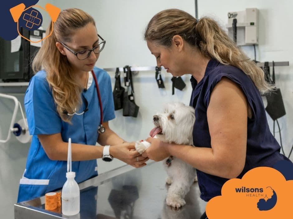
Talking with your vet: useful questions by phase
You don’t need to remember every cell type involved in healing. But having a mental map of the phases can help you ask focused questions.
Early days (inflammation + debridement)
You might ask:
“Does this look like normal post‑op inflammation, or are there signs of infection?”
“How much redness/swelling is acceptable, and for how long?”
“What type of debridement are we using—are you expecting the body to handle most of it, or will we need repeated cleaning at the clinic?”
“What should the bandage or wound look like day‑to‑day if things are going well?”
Middle phase (repair / proliferation)
You might ask:
“Are we seeing healthy granulation tissue?”
“Is the wound contracting and getting smaller at the pace you’d expect?”
“Would any advanced therapies (like PRF, stem cells, amniotic membrane, or laser) meaningfully change our outlook, or is standard care enough here?”
“How will we track progress—photos, measurements, rechecks?”
Later phase (maturation / remodeling)
You might ask:
“Even though it looks healed, how fragile is this area really?”
“How long should we protect it from rough play or certain activities?”
“Are there signs we should watch for that might suggest breakdown or recurrence?”
“Given my dog’s underlying conditions, are we at higher risk for future wounds?”
These conversations don’t make you a “difficult” client; they make you a well‑oriented partner in care.
What we know for sure—and what we’re still learning
Veterinary wound research is surprisingly rich in some areas and thin in others. It can help to know where the ground is solid and where it’s still shifting.
Well‑established
The four classic phases—inflammation, debridement, repair/proliferation, maturation/remodeling—occur in dogs much as they do in other mammals.[1][3][5][7][9][11][14]
These phases are overlapping, not strictly sequential.
Moist wound healing, appropriate debridement, and infection control significantly improve outcomes.[11][12][13]
Collagen remodeling during maturation increases wound strength to around 70–80% of original skin strength, but this can take months to years.[9][11]
Still emerging or uncertain
The optimal protocols for stem cell therapy and PRF: dosage, timing, delivery methods.[2][6]
The long‑term tissue quality after regenerative therapies vs. conventional healing.[2][4][6][8]
The cost‑benefit balance of advanced options for different kinds of wounds.
The best early markers to predict which wounds will become chronic or which dogs will struggle to heal.[2][4][6][8][11]
This uncertainty doesn’t mean these therapies are “experimental in a scary way,” but it does mean decisions often involve weighing promising evidence against cost, logistics, and your dog’s specific situation.
How understanding the phases can actually make this easier
Knowing the four phases isn’t about turning you into a vet. It’s about giving your brain a structure so that every new change in the wound doesn’t feel like chaos.
When the incision is red and warm on Day 2, you can think:“We’re in the inflammatory phase. I’ll watch for the boundaries of redness, but some of this is expected.”
When the wound looks pink and bumpy on Day 7, you can think:“This is granulation tissue in the repair phase. It’s supposed to look like it’s ‘filling in.’ I’ll confirm at our recheck.”
When the scar looks fine but your vet still recommends limiting rough play for a few more weeks, you can think:“We’re in the remodeling phase. It looks done, but the collagen is still organizing. We’re protecting the long game.”
That shift—from “What on earth is happening?” to “I know roughly where we are in the process”—is often what reduces the quiet background fear and self‑blame that can make wound care so emotionally draining.
Your dog’s body is doing something complex and ancient, one phase at a time. Your job is not to control that process, but to support it, ask clear questions, and protect the healing tissue as best you can. That’s already a lot—and it’s enough.
References
Raintree Veterinary Center. Dog Wound Care & Healing Stages.
Vilar JM, Batista M, Morales M, Santana M, Cuesta M, Arencibia A. Cutaneous wound healing in the dog: Allogeneic adipose-derived mesenchymal stem cell therapy. PLoS One.
Mooresville Animal Hospital. Dog Wound Care & Healing Stages.
Abd El-Aal AM, et al. Efficacy of xenogeneic fresh and lyophilized amniotic membranes on full-thickness skin wound healing in dogs. Scientific Reports (Nature).
Animal Hospital at Babcock Ranch. Dog Wound Care & Healing Stages.
De Rossi R, et al. Management of canine wounds using autologous platelet-rich fibrin (PRF). Veterinary and Comparative Orthopaedics and Traumatology. Wiley.
Pavletic MM. Principles of Wound Management and Healing in Small Animals. Vet Clin North Am Small Anim Pract. PMC/NIH.
Marchegiani A, et al. Photobiomodulation (Laser) Therapy for Incision Healing in Dogs. Frontiers in Veterinary Science.
PetVax. Dog Wound Healing Stages.
Central Pet Veterinary Clinic. Case Study on Open Wound Management.
Merck Veterinary Manual. General Principles of Wound Healing in Small Animals.
Swaim SF, Henderson RA. Moist Wound Healing in Small Animal Patients. Today’s Veterinary Practice.
DVM360. Wound Management Proceedings.
MSD Veterinary Manual. Stages of Wound Healing Table.


Comments